Left Shoulder Anatomy Diagram - Shoulder Rotator Cuff Tear Subscapularis Muscle Supraspinatus Muscle Broad Left Front Anatomy Acromion Scapula Png Pngwing / Starting with what is deepest, it goes:
Left Shoulder Anatomy Diagram - Shoulder Rotator Cuff Tear Subscapularis Muscle Supraspinatus Muscle Broad Left Front Anatomy Acromion Scapula Png Pngwing / Starting with what is deepest, it goes:. Click on a view to review its anatomy. Sechrest, md narrates an animated tutorial on the basic anatomy of the shoulder. Supraspinatus, infraspinatus, ters minor,.et), using interactive animations and labeled diagrams. Month ago i fell on my left shoulder while on a bush walk, hard fall. Mr is the best imaging modality to examen patients with shoulder pain and instability.
We added an horizontal menu at. Robin smithuis and henk jan van der woude. Did not undergo a x ray or. Just remember the articulating surfaces. Draw around to the other side, ghosting it as it goes around.
Left Arm Muscle Model Labeled Google Search Body Anatomy Arm Muscle Anatomy Human Body Anatomy from i.pinimg.com Choose from 500 different sets of flashcards about shoulder anatomy on quizlet. Did not undergo a x ray or. Learn their origins/insertions, functions & exercises. We added an horizontal menu at. Starting with what is deepest, it goes: This webpage presents the anatomical structures found on shoulder mri. Click on a view to review its anatomy. In this episode we'll go over the simple structure and the anatomical details of both the clavicle and scapula.
Your email address will not be published.
Required fields are marked *. Human anatomical atlas of the shoulder : Your email address will not be published. Just remember the articulating surfaces. Click on a view to review its anatomy. Tutorials on the shoulder muscles (e.g rotator cuff muscles: The transverse humeral ligament is not shown on this diagram. Left shoulder by genusfotografen / wikimedia. Principles of anatomy and physiology [with a brief atlas of the skeleton, s. Did not undergo a x ray or. The rotator cuff is an anatomical term given to the group of four muscles and their tendons that act to stabilize the the left shoulder and acromioclavicular joints, and the proper ligaments of the scapula. You can see it enclosing the glenohumeral joint and you can see its attachment on the anatomical neck that's the shoulder joint. In this episode of eorthopodtv, orthopaedic surgeon randale c.
They're the muscles you see when you roll up. This is because the deltoids are what you would consider the major muscles of the shoulder anatomy; Learn their origins/insertions, functions & exercises. Did not undergo a x ray or. Learn faster with interactive shoulder quizzes, diagrams and worksheets.
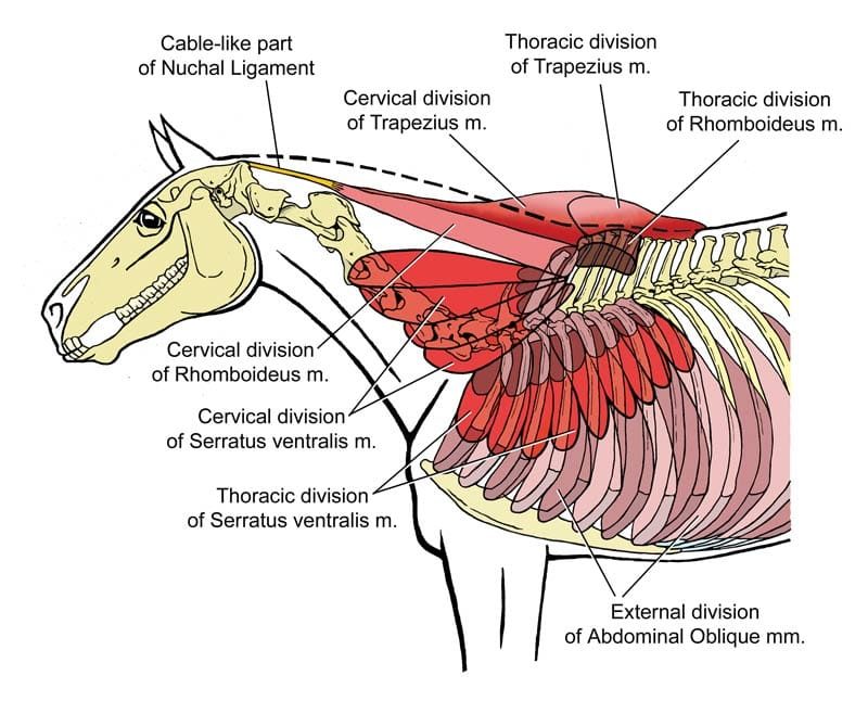
Equine Reciprocating Systems Examining The Shoulder To Thorax Junction American Farriers Journal from www.americanfarriers.com Movements of the human shoulder represent the result of a complex dynamic interplay of structural bony anatomy and biomechanics, static ligamentous and tendinous restraints, and dynamic muscle forces. This acts as the bony framework by which the muscles of the chest, upper back and shoulder connect the upper limb to the trunk of the body and control it's movements.the clavicle connects to the sternum via the. Learn their origins/insertions, functions & exercises. Related online courses on physioplus. Draw around to the other side, ghosting it as it goes around. The rotator cuff is an anatomical term given to the group of four muscles and their tendons that act to stabilize the the left shoulder and acromioclavicular joints, and the proper ligaments of the scapula. 7 draw labelled diagram showing the relations of shoulder joint. Bone, then ligaments of the joint capsule, with tendons and muscles on top.
Just remember the articulating surfaces.
Left shoulder by genusfotografen / wikimedia. Draw around to the other side, ghosting it as it goes around. This webpage presents the anatomical structures found on shoulder mri. Supraspinatus, infraspinatus, ters minor,.et), using interactive animations and labeled diagrams. Editor · aug 6, 2017 ·. Constituents of the shoulder joint. Learn their origins/insertions, functions & exercises. This acts as the bony framework by which the muscles of the chest, upper back and shoulder connect the upper limb to the trunk of the body and control it's movements.the clavicle connects to the sternum via the. We think this is the most useful anatomy picture that you need. Human anatomical atlas of the shoulder : To keep things simple, we can divide the shoulder into layers. Related online courses on physioplus. Learn about shoulder anatomy with free interactive flashcards.
Leave a reply cancel reply. 7 draw labelled diagram showing the relations of shoulder joint. Your email address will not be published. Webmd's shoulder anatomy page provides an image of the parts of the shoulder and describes its function, shoulder problems, and more. Learn their origins/insertions, functions & exercises.
Shoulder from www.daviddarling.info Learn faster with interactive shoulder quizzes, diagrams and worksheets. Want to learn more about it? Learn their origins/insertions, functions & exercises. Understanding how the different layers of the shoulder are built and connected can help you understand how the shoulder works, how it can be injured, and how challenging recovery can be. Mr is the best imaging modality to examen patients with shoulder pain and instability. Axial slice of t1 weighted mri with all anatomical structures labeled. As a ball and socket synovial. Leave a reply cancel reply.
Human anatomical atlas of the shoulder :
This acts as the bony framework by which the muscles of the chest, upper back and shoulder connect the upper limb to the trunk of the body and control it's movements.the clavicle connects to the sternum via the. Robin smithuis and henk jan van der woude. Sobotta atlas of anatomy general anatomy and musculoskeletal system jens waschke|friedrich. This is because the deltoids are what you would consider the major muscles of the shoulder anatomy; The shoulder joint is the connection between the chest and the upper extremity. Did not undergo a x ray or. The shoulder is a complex combination of bones and joints where many muscles act to provide the widest range of motion numerous muscles help stabilize the three joints of the shoulder while giving it motion. Click on a view to review its anatomy. Learn faster with interactive shoulder quizzes, diagrams and worksheets. Learn all about the anatomy and function of the shoulder girdle fast and efficiently in this article shoulder girdle : Then, draw the shoulder girdle on top of the ribcage. Axial slice of t1 weighted mri with all anatomical structures labeled. Human anatomical atlas of the shoulder :
Use the mouse scroll wheel to move the images up and down alternatively use the tiny arrows (>>) on both side of the image to move the images shoulder anatomy diagram. We think this is the most useful anatomy picture that you need.